RESEARCH
# Research at MAS Lab
MAS Lab develops multimodal acoustic systems that connect radiation, light, and ultrasound to quantitative imaging, disease monitoring, and therapeutic stimulation. Our central engineering question is: how can deposited energy or acoustic stimulation be converted into reliable, interpretable, and quantitative biomedical information?
MAS Lab의 연구는 방사선·광·초음파를 공통된 음향 시스템 관점에서 연결합니다. 에너지 침적 또는 초음파 자극에서 시작해 신호 발생·검출, 재구성, 정량화, 생체 해석으로 이어지는 전체 파이프라인을 설계합니다. AI와 computational imaging은 각 연구축을 연결하고 정량성을 높이는 핵심 도구로 활용합니다.
01 · RADIATION ACOUSTICS
Radiation-Induced Acoustic Imaging & Dosimetry
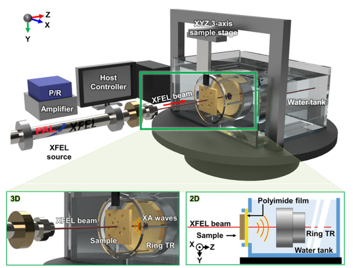
We investigate acoustic waves generated by ionizing-radiation energy deposition. Our goals are to develop sensitive radiation-acoustic detection systems, reconstruct energy-deposition distributions, and establish quantitative links between acoustic signals and radiation dose. Topics include X-ray and radiotherapy-induced acoustics, real-time dosimetry, system synchronization, physics-based simulation, low-SNR reconstruction, and extensions toward emerging radiation sources.
학생 연구 예시: 방사선-음향 신호 검출, X-ray/radiotherapy 실험, 선량-음향 신호 calibration, Monte Carlo/음향 시뮬레이션, 저 SNR 신호 향상, 3D 선량 영상화.
02 · ACOUSTIC BIOIMAGING
Photoacoustic & Ultrasound Imaging
We develop photoacoustic and ultrasound platforms for structural, functional, and molecular imaging. Research spans 3D volumetric imaging, high-speed acquisition, spectral imaging, multimodal integration, image reconstruction, and system optimization for preclinical and translational applications.
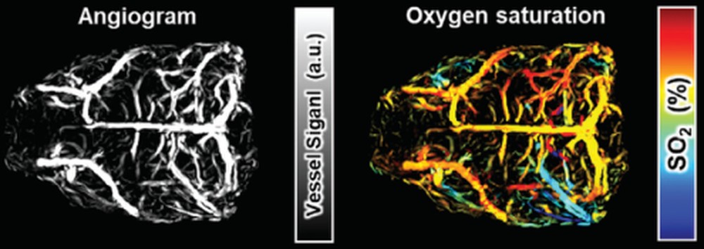
학생 연구 예시: 광음향/초음파 시스템 제작, 3D 재구성, 혈관 영상화, 산소포화도 분석, 고속 스캐닝, 멀티모달 영상, 영상 품질 및 시스템 최적화.
03 · QUANTITATIVE BIOMARKERS
Disease Monitoring & Quantitative Biomarkers
We use longitudinal imaging to quantify biological change rather than only produce images. We study cerebrovascular and fluid dynamics, tumor microenvironments, oxygenation, pharmacokinetics, treatment response, and recovery, with the goal of deriving repeatable and interpretable imaging biomarkers.
응용 방향: 뇌혈관·뇌척수액, 종양 미세환경, 약물·조영제 biodynamics, 질환 진행, 치료 반응을 반복 측정하고 vascular, oxygenation, molecular, fluid-dynamic 지표를 정량 바이오마커로 정의·검증합니다.
04 · ACOUSTIC INTERVENTION
Ultrasound Stimulation & Therapy
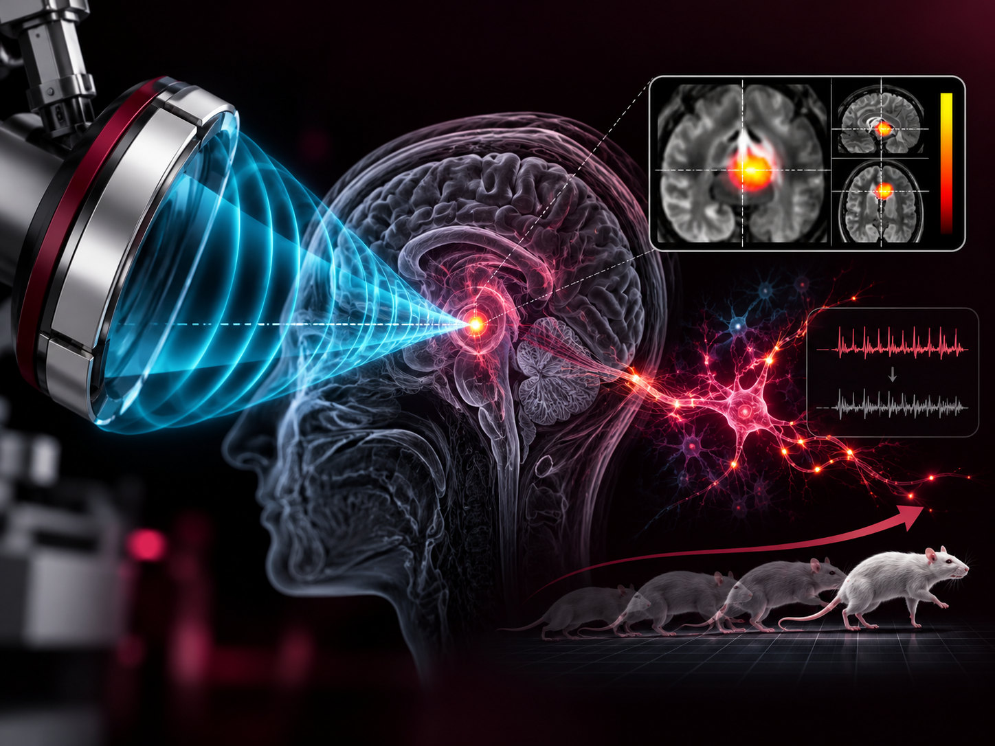
We engineer ultrasound exposure and monitoring strategies for neuromodulation, therapeutic stimulation, and rehabilitation-oriented applications. Rather than treating stimulation as a black box, we aim to systematically optimize frequency, pressure, pulse structure, and exposure conditions, while quantifying the resulting biological response with imaging and physiological measurements.
학생 연구 예시: 초음파 주파수·강도·펄스 조건 최적화, 신경자극 및 therapeutic ultrasound 실험, 자극 전후 생체 반응 영상화, 재활 관련 기능 변화의 정량 평가, image-guided stimulation.
CROSS-CUTTING METHODS
AI & Computational Imaging
AI is not treated as a separate research destination in MAS Lab. Instead, model-based reconstruction, deep learning, physics-informed learning, segmentation, multimodal registration, uncertainty estimation, and GPU computing are used across all four research directions to improve sensitivity, speed, robustness, and quantitative reliability.
AI는 독립적인 연구 목적이라기보다 방사선 유도 음향영상, 광음향·초음파 영상, 바이오마커 정량화, 초음파 자극 연구 전반을 고도화하는 공통 방법론으로 활용합니다. 물리 모델과 AI를 결합하여 저 SNR, 제한 시야, inverse problem, 자동 정량화 문제를 해결합니다.
REPRESENTATIVE WORK
Selected Research Highlights
The images below are reproduced or adapted from representative publications and are linked to the original articles.
RADIATION ACOUSTICS · 2024
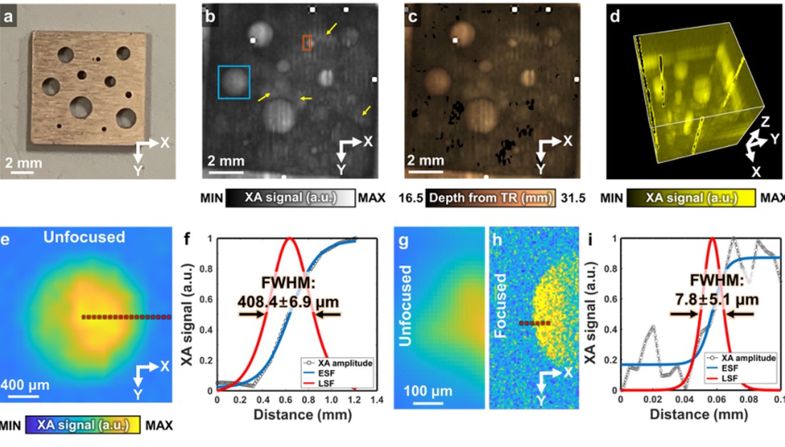
X-ray free-electron laser induced acoustic microscopy
XFEL excitation enables micron-scale X-ray-induced acoustic microscopy and demonstrates the feasibility of a new X-ray acoustic regime.
Photoacoustics ↗
Figure reproduced from Choi et al., Photoacoustics 35, 100587 (2024), CC BY-NC-ND 4.0.
RADIATION ACOUSTICS · 2022
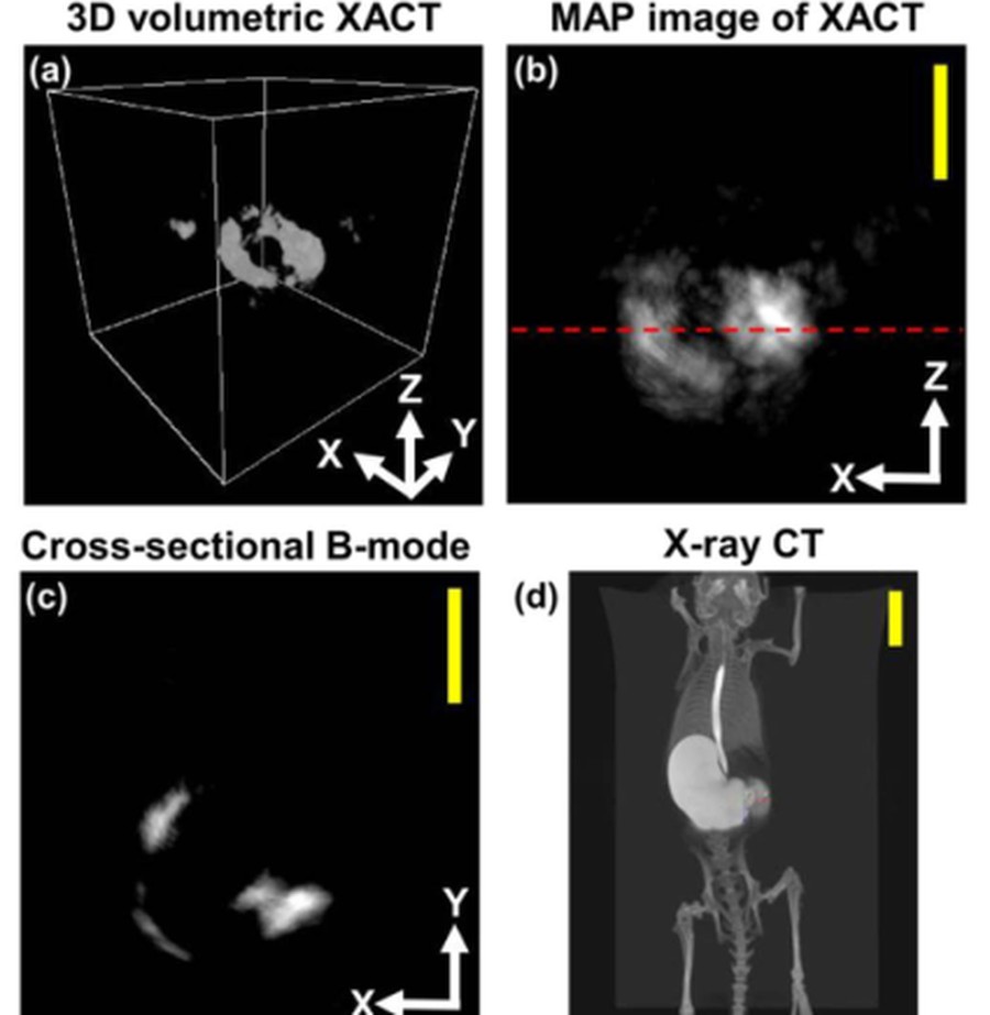
In situ X-ray-induced acoustic computed tomography
Contrast-enhanced XACT demonstrates volumetric in situ small-animal imaging and establishes a route toward biomedical X-ray acoustic applications.
Optics Letters ↗
Figure reproduced from Choi et al., Optics Letters 47, 90-93 (2022).
BIOAPPLICATION · 2026
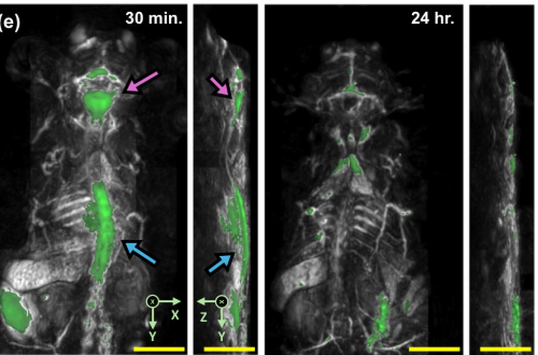
Photoacoustic monitoring of CSF dynamics and glymphatic function
Whole-body PACT tracks CSF transport, anesthesia-dependent efflux, and impaired clearance in an Alzheimer’s disease mouse model.
Nature Communications ↗
Figure adapted from Choi et al., Nature Communications 17, 2677 (2026), CC BY 4.0.
NEUROIMAGING · 2025
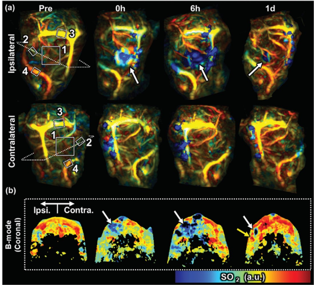
Early-stage ischemic stroke monitoring
Whole-brain PACT visualizes cerebrovascular remodeling and vessel-specific oxygenation changes following photothrombotic stroke.
Advanced Science ↗
Figure adapted from Kim et al., Advanced Science 12, 2409361 (2025), CC BY 4.0.
IMAGING SYSTEM · 2025
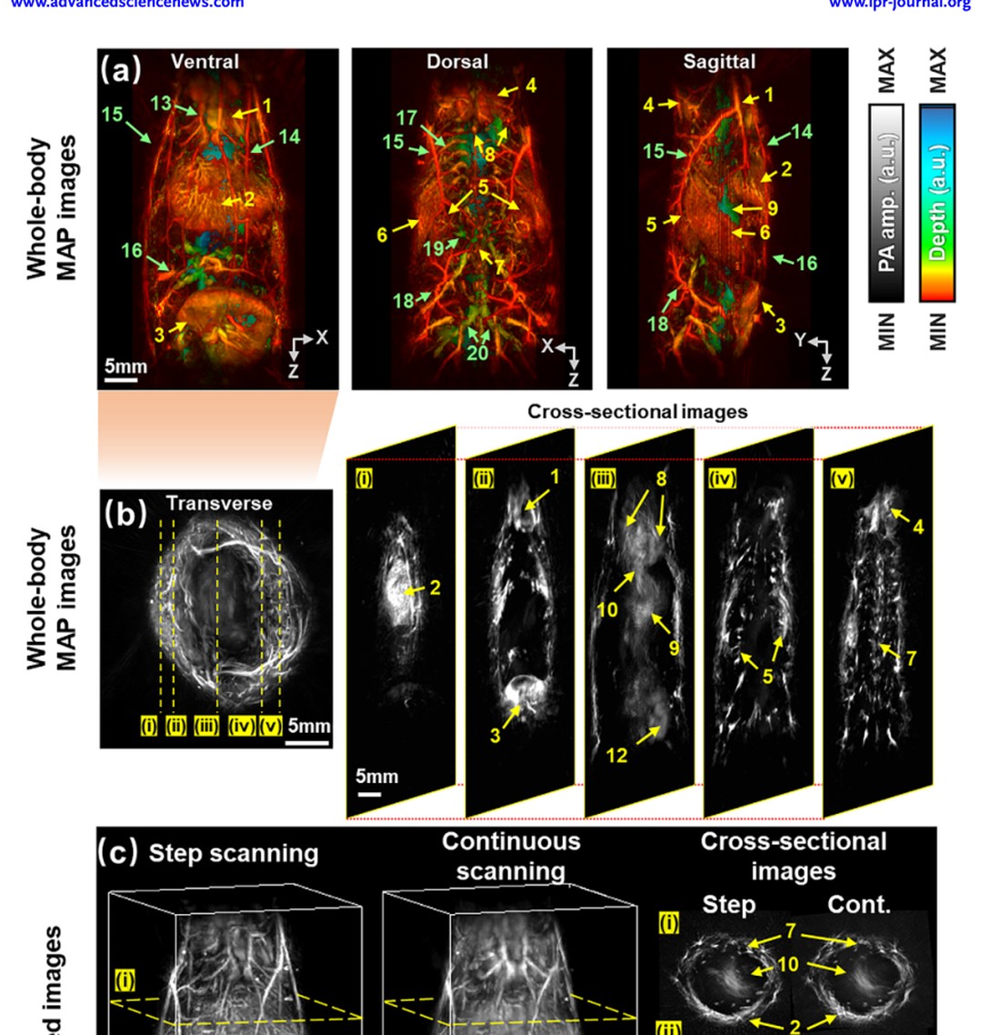
Multiplane spectroscopic whole-body PACT
Continuous rotary scanning extends tomographic coverage for high-resolution structural, functional, and pharmacokinetic small-animal imaging.
Laser & Photonics Reviews ↗
Wiley Top Viewed Article 2025
Figure reproduced from Yang et al., Laser & Photonics Reviews 19, 2400672 (2025), CC BY-NC-ND 4.0.
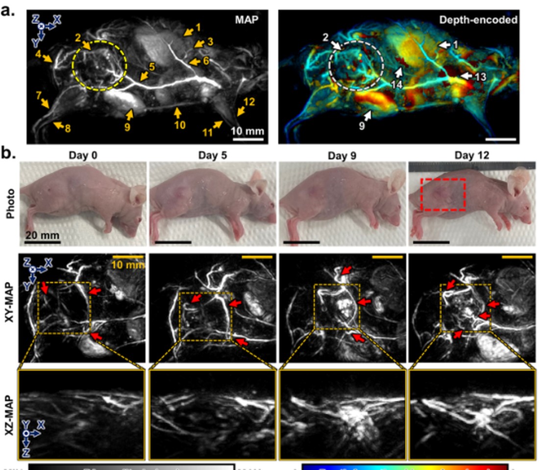
ONCOLOGY · 2024
3D multiparametric PACT of primary and metastatic tumors
Longitudinal structural, oxygenation, and pharmacokinetic measurements characterize tumor progression and metastasis in living mice.
ACS Nano ↗
Figure reproduced from Kim et al., ACS Nano 18, 18176-18190 (2024), CC BY-NC-ND 4.0.
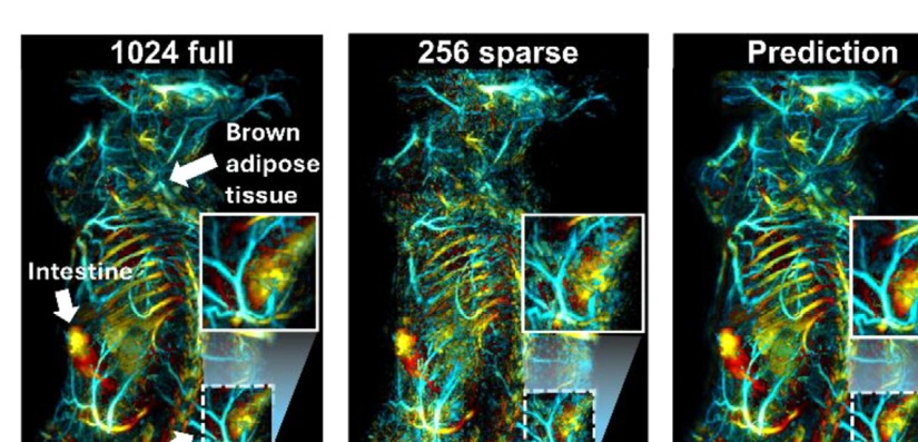
COMPUTATIONAL IMAGING · 2026
Hybrid diffusion model for multiparametric 3D PACT
Deep generative reconstruction restores structural and functional information from substantially reduced ultrasound-element sampling.
Advanced Science ↗
Figure adapted from Jeong et al., Advanced Science 13, e13624 (2026), CC BY 4.0.
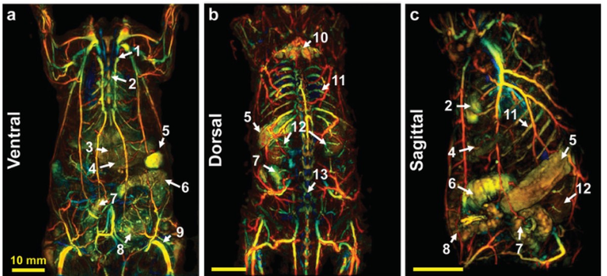
COMPUTATIONAL IMAGING · 2023
Deep learning enhanced multiparametric dynamic volumetric PACT
A 3D progressive U-Net recovers limited-view volumetric information and enables high-speed structural, functional, and contrast-enhanced imaging.
Advanced Science ↗
Back CoverWiley Top Downloaded Article
Figure adapted from Choi et al., Advanced Science 10, 2202089 (2023), CC BY 4.0.
COLLABORATIVE RESEARCH
Research Collaborators & Networks
MAS Lab grows through project-level collaborations across biomedical imaging, radiology, nuclear medicine, nanotechnology, neuroscience, rehabilitation, and translational science.
국내외 연구진과의 공동연구를 통해 방사선·음향 계측, 영상 시스템 개발, computational imaging, 전임상·임상 검증, 자극·치료 연구까지 범위를 확장합니다.
Institution names indicate research relationships or networks at the project/researcher level and do not imply institutional endorsement.